
Updated on: November 20, 2026
Originally published on: November 20, 2026
Ever wonder how your dentist knows exactly where to put a dental implant when so much of the work happens underneath your gums? That’s where digital imaging for dental implants comes in. A 3D scan lets your dentist see the jawbone beneath that gap in your smile, while a small handheld scanner captures the shape of the teeth and gums around it. Put those two pictures together, and your dentist can plan where the implant will go and how your new tooth will fit alongside the others.
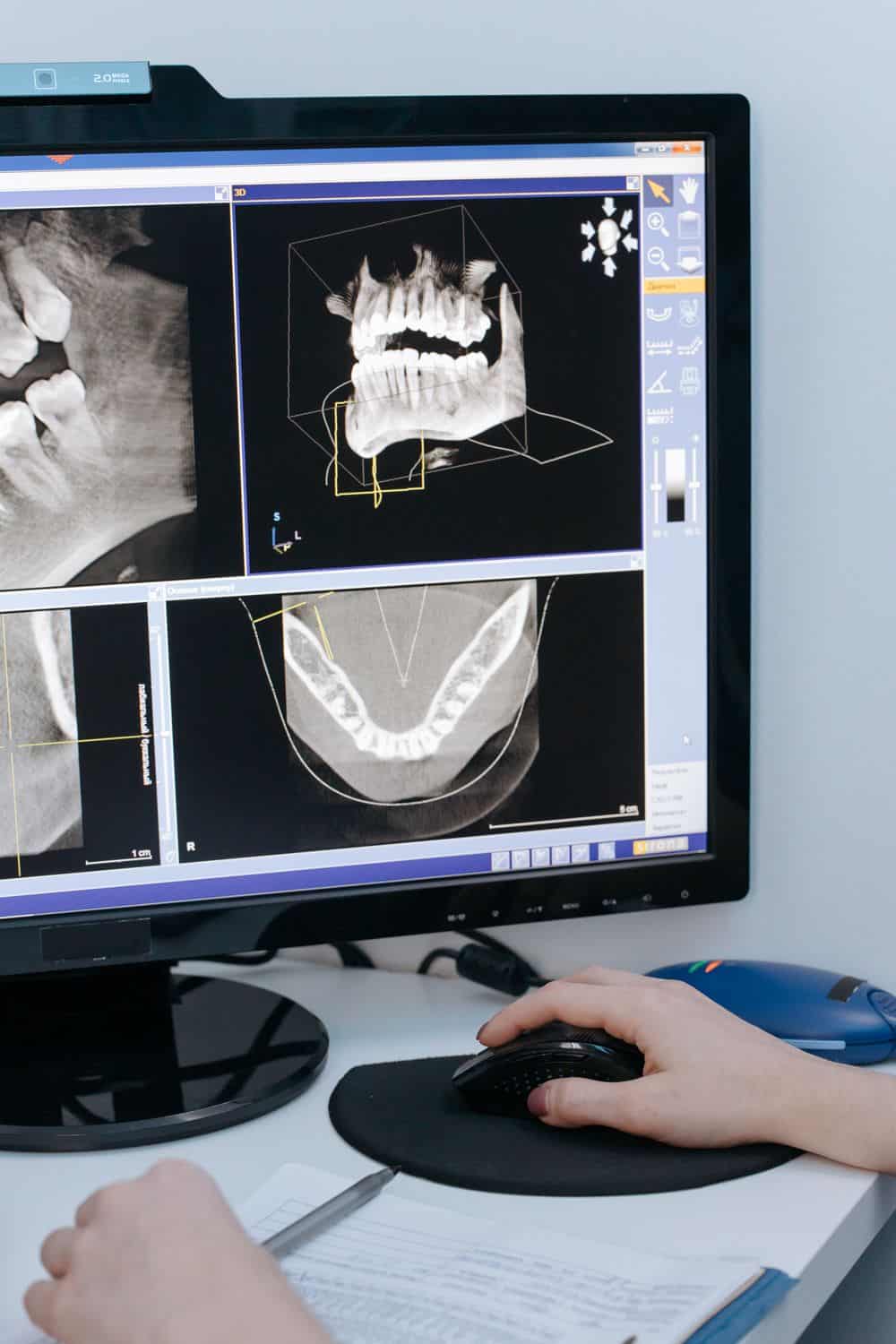
Losing a permanent tooth changes how you chew, speak, and feel about your appearance. For decades, replacing that missing tooth meant enduring trays of goopy impression material and relying on flat, 2-dimensional X-rays that forced your dentist to estimate your underlying bone structure.
Modern digital imaging eliminates that uncertainty right from your very first consultation. By combining 3D cone-beam computed tomography (CBCT) with optical intraoral scanners, your restorative dentist maps your entire oral anatomy down to the sub-millimeter before treatment even begins.
How Do Virtual Surgical Guides Improve Placement and Aesthetics?
Once your dentist merges your optical surface scan with your 3D bone map, your entire mouth exists as a virtual surgical playground. Rather than waiting until the day of your procedure to determine the ideal angle or depth for a restoration, your care team performs the surgery digitally first. This virtual rehearsal transforms complex restorative dentistry into a predictable, streamlined experience.
During this planning phase, your restorative dentist evaluates your bone readiness and designs a customized treatment blueprint. When clinicians place permanent Dental Implants using 3D diagnostic data, they align the titanium post with the exact position of your future crown. Working backward from the final tooth appearance guarantees that the underlying support structure emerges through your gums at the most natural angle possible.
Turning Digital Blueprints Into Physical Guides
After finalizing the virtual plan, your dental team sends the digital file to a 3D printer or milling unit to fabricate a custom surgical guide. This clear, medical-grade resin stent fits snugly over your existing teeth during your appointment, featuring precision-engineered metal sleeves that direct the surgical instruments.
Key Advantages of Guided Placement
- Pinpoint depth and angle control: The guide physically prevents the instruments from drifting off course or going deeper than planned.
- Minimally invasive incisions: Because your dentist already knows where the bone is thickest, keyhole access often replaces large gum flaps.
- Shorter chair time: Pre-planned trajectories eliminate mid-procedure adjustments, letting you get back to your day faster.
- Smoother post-op recovery: Less tissue disruption typically leads to minimal swelling, reduced discomfort, and quicker healing.
Custom Abutments for Natural-Looking Crowns
Connecting a titanium post to a visible porcelain crown requires an intermediate piece called an abutment. Stock, off-the-shelf abutments come in generic shapes that can sometimes leave awkward gaps near the gumline or feel bulky against your tongue. Digital workflows allow dental laboratories to mill custom titanium or zirconia abutments shaped specifically to match your unique gum contours.
Your dentist also uses the digital bite registration from your intraoral scan to check how your upper and lower teeth meet when you chew. By analyzing occlusal forces on screen, the lab shapes the final crown so it shares biting pressure evenly with your neighboring teeth. Balanced bite forces protect your new restoration from premature wear and keep your jaw joints comfortable.
How Does 3D Digital Imaging Map Your Mouth Without Messy Molds?
Traditional dental impressions require biting down on a tray filled with cold, putty-like alginate or polyvinyl siloxane for several minutes. If you have a strong gag reflex or sensitive teeth, that process alone can make you dread the dental chair.
Digital intraoral scanners replace those physical trays with a sleek, handheld optical wand that captures thousands of high-definition photos per second.
As your dentist glides the wand over your teeth and gums, specialized software stitches those images together into a live, full-color 3D model on a chairside monitor. You get to see what your clinician sees in real time, which makes understanding your treatment options much easier. According to DDS News, a quick 2-minute digital scan improves patient communication so much that treatment acceptance jumped from 50% in 2018 to 80% today.
Looking Beneath the Gumline With CBCT Scans
Surface scans show the visible contours of your teeth and soft tissue, while a CBCT machine looks deep beneath the gums. Unlike standard medical CT scans that require lying inside a closed tunnel, a dental CBCT unit rotates quietly around your head in under 20 seconds while you sit or stand comfortably. This single rotation captures hundreds of cross-sectional angles to build a complete 3D map of your jaw.
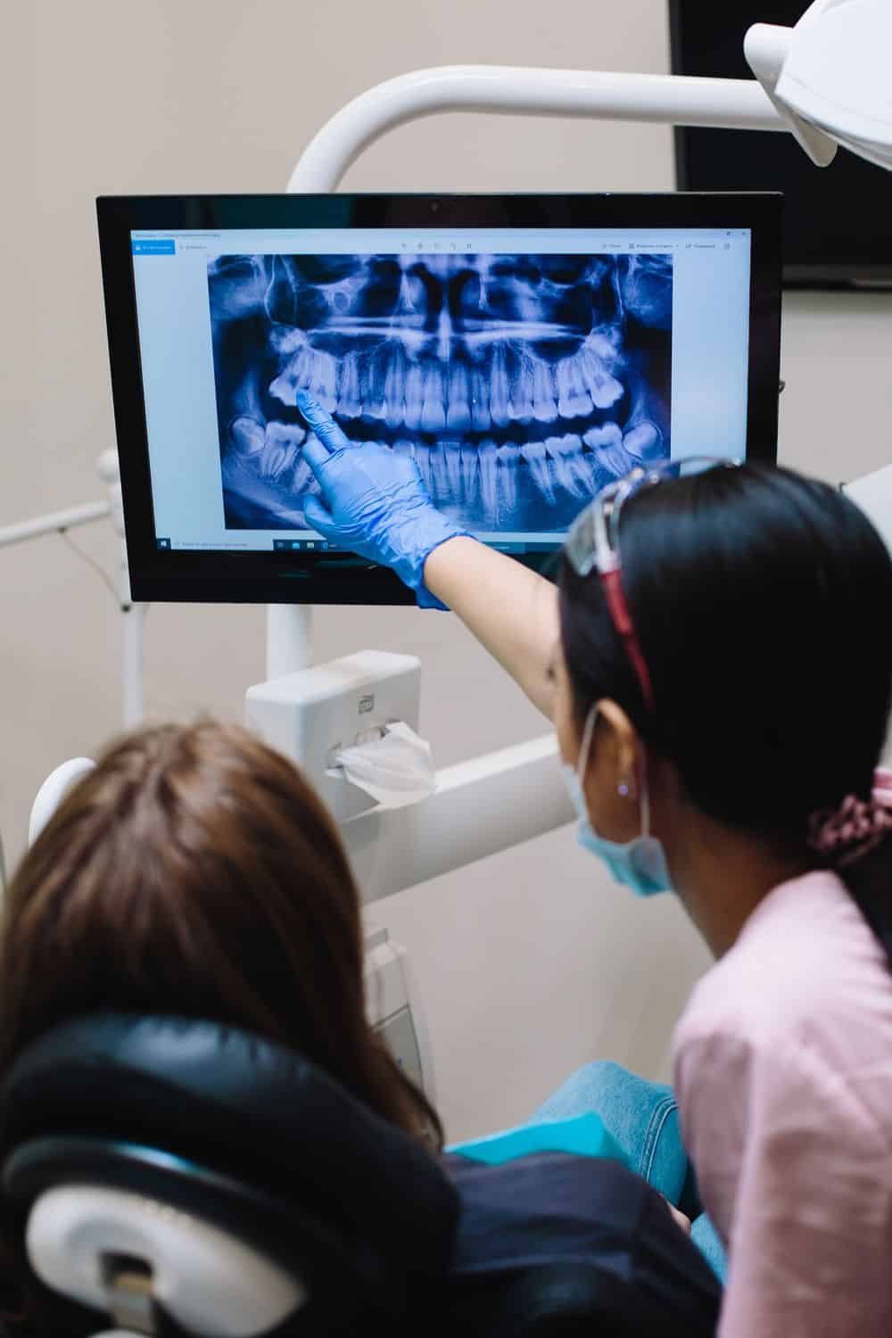
What Your Dentist Sees in a 3D Scan
With this volumetric data on screen, your clinician inspects critical anatomical structures from every possible angle before making a single incision.
- Jawbone volume and density: Your dentist measures the exact height, width, and thickness of your jawbone to confirm it can anchor a permanent restoration.
- Sinus cavity boundaries: For upper back teeth, the scan reveals the exact floor of your maxillary sinus so your dentist can avoid accidental perforation.
- Nerve pathway locations: In the lower jaw, the 3D view pinpoints the inferior alveolar nerve canal, keeping surgical placement at a safe distance.
- Hidden root infections: High-resolution cross-sections expose silent cysts, cracked roots, or lingering bone loss around neighboring teeth that flat X-rays often miss.
Keeping Radiation Exposure Low
You might wonder if a 3D scan exposes you to excessive radiation compared to traditional dental films. Modern CBCT systems use focused, cone-shaped beams that target only the specific area of your jaw under evaluation.
As reported by DentistryIQ, updated clinical guidelines emphasize the ALADAIP principle, ensuring radiation doses stay as low as diagnostically acceptable while artificial intelligence assists clinicians in interpreting scans with greater consistency.
Why Choose Digitally Planned Restorations Over Bridges or Dentures?
When weighing your options for replacing missing teeth, you generally choose between removable dentures, traditional fixed bridges, or permanent titanium restorations. While dentures and bridges offer shorter upfront timelines, they sit above the gumline and do not interact with your underlying jawbone. Understanding how each option affects your long-term oral health helps you make a confident investment in your smile.
Preserving Your Jawbone Over Time
Your natural tooth roots stimulate your jawbone every time you bite down, signaling your body to keep sending nutrients and minerals to that area. When a tooth goes missing, that stimulation stops, and the surrounding bone begins to shrink through a natural process called resorption.
Removable dentures rest directly on the gums and can actually accelerate bone loss through daily friction, eventually causing the appliance to slip or click when you talk. Traditional dental bridges span the gap by anchoring onto the two healthy teeth on either side of the missing space.
Stepping into a modern dental office equipped with 3D imaging changes how you participate in your own care. Instead of guessing how a procedure might turn out, you can review a clear visual roadmap tailored to your exact anatomy.
Ask your dentist whether they combine CBCT scans with digital intraoral impressions to plan permanent restorations, and request to see your 3D bone model on screen. Pairing advanced diagnostic imaging with guided placement gives you a safer procedure, a more comfortable recovery, and a custom tooth replacement built to feel like your own.